pubmed: 25895010 doi: 10.1021/acs.est.5b00182 issn: 1520-5851 issn: 0013-936x
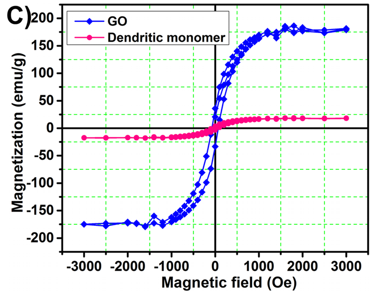
"Figure 1. Characterization of dendritic monomer by ... (C) VSM techniques"

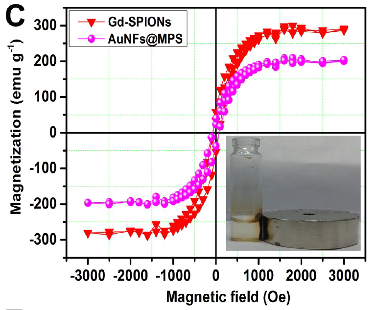
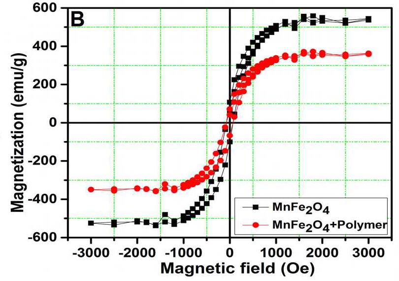
Hysteresis data points have elsewhere appeared as "Magnetic hysteresis loop of Gd-SPIONs and AuNFs@MPS"

"Figure 2. FE-SEM image of (A) GO, (B) GO/silane@MNPs; (C) FE-SEM, (D) TEM, and (E) HR-TEM images of dendritic monomer; (F) FT-IR spectra of (a) Eu (III)-imprinted polymer, (b) after rebinding, and (c) extraction."

The three spectra in 2F are indistinguishable from three copies of the same line, with vertical offsets and a horizontal offset for spectrum (b).
Figure 2D -- compare with inset, showing nanohybrid, in "Figure 1: ... (F) nanohybrid, inset showing the magnified TEM image." from https://pubpeer.com/publications/C94874C49B2479968F262F83C9F128#3

Attach files by dragging & dropping,
selecting them, or pasting
from the clipboard.
![]() Uploading your files…
We don’t support that file type.
with
a PNG, GIF, or JPG.
Yowza, that’s a big file.
with
a file smaller than 1MB.
This file is empty.
with
a file that’s not empty.
Something went really wrong, and we can’t process that file.
Uploading your files…
We don’t support that file type.
with
a PNG, GIF, or JPG.
Yowza, that’s a big file.
with
a file smaller than 1MB.
This file is empty.
with
a file that’s not empty.
Something went really wrong, and we can’t process that file.
