doi: 10.1016/j.biopsych.2009.09.031 issn: 0006-3223 pubmed: 19932469 issn: 1873-2402
 ,
Kalindi Bakshi
,
Changpeng Shen
,
Maya Frankfurt
,
Caryn Trocmé-Thibierge
,
Philippe Morain
,
Kalindi Bakshi
,
Changpeng Shen
,
Maya Frankfurt
,
Caryn Trocmé-Thibierge
,
Philippe Morain
A similarity of a Figure 1A Actin blot with an Actin blot in Figure 12A of Wang et al., Neuroscience (2005) 135: 247-261 , DOI: 10.1016/j.neuroscience.2005.06.003 was discussed in a letter to the FDA found here: https://www.regulations.gov/docket/FDA-2021-P-0930/document
Here is a copy of the comparison of the two Actin blots (not my own finding, but I agree with the similarity).
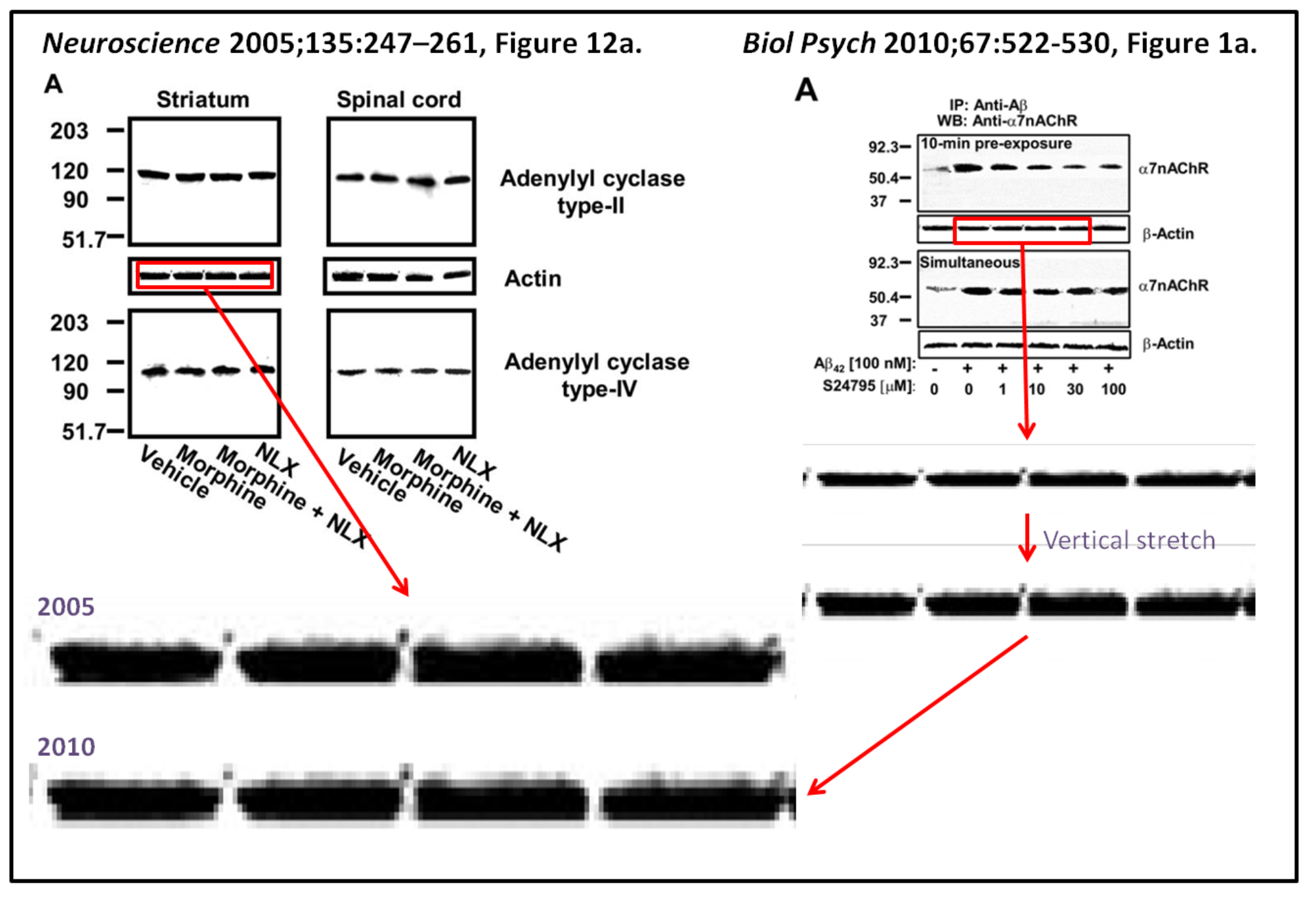
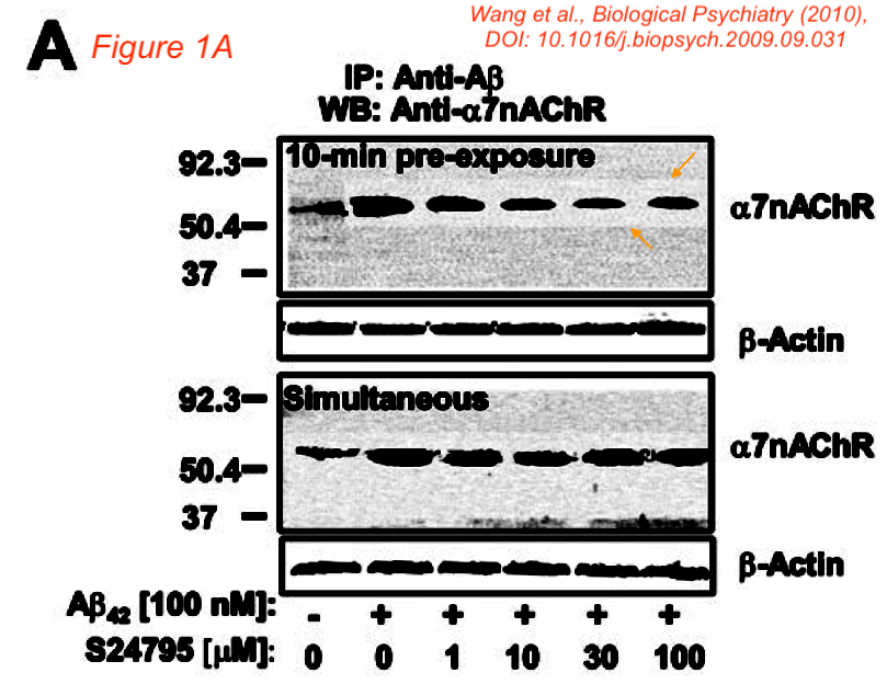
In addition - not mentioned in the report to the FDA - Figure 1A appears to show a lighter background around some, but not all bands in the top a7nAChR blot. A similar lighter area is visible around all bands in the lower blot.
Image made darker to bring out details.
Might the authors still have the original blots to take away any concerns?


An expression of concern was issued for this article, it reads:
The Journal is issuing an expression of concern for the article, “S 24795 Limits β-Amyloid–α7 Nicotinic Receptor Interaction and Reduces Alzheimer's Disease-Like Pathologies”. The editors are aware of concerns about Western blots in this article. These and other concerns are currently under investigation by the academic authorities at the City University of New York (CUNY). The Journal is awaiting the outcome of that investigation before taking action.

Link to Expression of Concern, issued 18 June 2024: https://www.biologicalpsychiatryjournal.com/article/S0006-3223(24)01386-6/fulltext
Attach files by dragging & dropping,
selecting them, or pasting
from the clipboard.
![]() Uploading your files…
We don’t support that file type.
with
a PNG, GIF, or JPG.
Yowza, that’s a big file.
with
a file smaller than 1MB.
This file is empty.
with
a file that’s not empty.
Something went really wrong, and we can’t process that file.
Uploading your files…
We don’t support that file type.
with
a PNG, GIF, or JPG.
Yowza, that’s a big file.
with
a file smaller than 1MB.
This file is empty.
with
a file that’s not empty.
Something went really wrong, and we can’t process that file.